| |
Título: Arial 14 puntos, negritas, mayúsculas.
Autores: Nombre, apellido de cada autor , marcando al autor corresponsal con *. Teléfono y el e-mail del autor corresponsal . Arial 12 puntos, negritas.
Direcciones: Departmento, Institución, Ciudad, Código postal, país. Arial 12 puntos.
Antecedentes: Arial, 12 puntos.
Objetivos: Arial, 12 puntos.
Materiales y Métodos: Arial, 12 puntos.
Resultados y Discusión: Arial, 12 puntos.
Conclusiones: Arial, 12 puntos.
Referencias: Arial, 12 puntos. |
|
| |
|
COMPARATIVE METHODOLOGY FOR THE QUANTIFICATION AND IDENTIFICATION
OF AFLATOXINS IN LIVER OF LAYING HENS.
Mariana Díaz-Zaragoza, Magda Carvajal*, Ignacio Méndez, Nahlleli Civi-Chilpa,
Ernesto Ávila- González, César Flores.
Universidad Nacional Autónoma de México, Ciudad Universitaria, Coyoacán, 04510 México, D,F.
*Tel: 5255+ 5622-9138 magdac@servidor.unam.mx
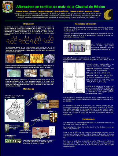
Background:Aflatoxins (AF) are secondary metabolites produced by strains of the fungi Aspergillus flavus, A. parasiticus and A. nomius, and they turn out to be a natural and dangerous contaminant of several cereal grains such as maize and sorghum. They are a great risk for both human and animal health. There are four important AF: AFB1, AFB2, AFG1 and AFG2, from which AFB1 is the most toxic, and it is considered a potent mutagen and carcinogen. AF are the most important mycotoxins in the broiler industry because they cause great economic losses, when poultry are fed with contaminated grains.
Aim: To quantify and identify AF in liver of laying hen, and to determine the best chemical method for their AF detection.
Materials and Methods: A total of 25 laying hens of 121-week old in a second cycle of egg production, were distributed at random in three experimental groups and placed in individual cages. The control group had 9 hens fed with 2 kg of milled and homogenized clean sorghum, without AFB1, every day for one week; and of two treated groups of 8 hens each, fed with contaminated sorghum with a low (30 µg/kg) or a high (500 µg/kg) AF concentration. After a week, the hens were killed by cervical dislocation and their livers were removed. The extraction method for AF was the one proposed by Qian & Yang (1984), and the chemical method with immunoaffinity columns given by Koeltzow & Tanner (1990). The quantification of AF was achieved by high-performance liquid chromatography (HPLC). The liver was the tissue with higher contamination and number of identified AF.
Results and Discussion: The livers had AFB1 (0.7-9.7 µg/kg), AFB2 (0.06-0.6 µg/kg), AFG1 (78.7-121.0 µg/kg), AFG2 (16.2-18.7 µg/kg), AFM1 (10.3-591.8 µg/kg), AFP1 (96.2-771.3 µg/kg) and AFL (4.8-12.6 µg/kg), from which AFG1, AFG2, AFM1 and AFP1 were obtained in greater amount. The determination of the appropriate chemical method for the quantification of AF settled down when the percentage of recovery of both methods was made. In both chemical methods around 80% of recovery was obtained which is an acceptable recovery. Nevertheless, the method of Qian & Yang (1984) had a lower limit of detection (0.5 ng/mL), while the limit of detection in the chemical method with immunoaffinity columns was of 5 ng/mL for liver.
Conclusion: In conclusion, the best chemical method for the quantification of AF, was the proposed by Qian & Yang (1984), however, it requires a modification to obtain a recovery of 80 to 90%, which consists of the addition of both AF elutions, from the first and second Supelclean LC-18 SPE columns.
References:
Qian, G-S & Yang, G. C. 1984. Rapid extraction and detection of aflatoxins B1 and M1 in beef liver. Journal Agricultural and Food Chemistry, 32:1071-1073.
Koeltzow, D. E. & Tanner, S. N. 1990. Comparative evaluation of commercially available test methods. Journal Association of Official Analytical Chemists, 73: 584-589.
|
|
|